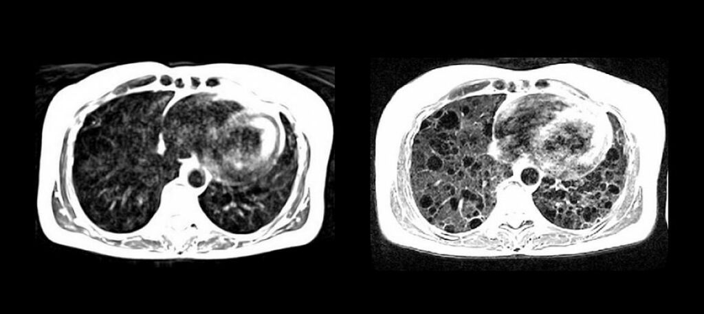
National Institutes of Health researchers, along with researchers at Siemens, have developed a high-performance, low magnetic-field MRI system that vastly improves image quality of the lungs and other internal structures of the human body. The new system is more compatible with interventional devices that could greatly enhance image-guided procedures that diagnose and treat disease, and the system makes medical imaging more affordable and accessible for patients.
The low-field MRI system may also be safer for patients with pacemakers or defibrillators, quieter, and easier to maintain and install. The study, funded by the National Heart, Lung, and Blood Institute (NHLBI), part of the National Institutes of Health, appears today in the journal Radiology.
The trend in recent years has been to develop MRI systems with higher magnetic field strengths to produce clearer images of the brain. But, researchers calculated that using those same state-of-the-art systems — at a modified strength — might offer high quality imaging of the heart and lungs. They found that metal devices such as interventional cardiology tools that were once at risk of heating with the high-field system were now safe for real-time, image-guided procedures such as heart catherization.
“We continue to explore how MRI can be optimized for diagnostic and therapeutic applications,” said Robert Balaban, Ph.D., scientific director of the Division of Intramural Research and chief of the Laboratory of Cardiac Energetics at NHLBI. “The system reduces the risk of heating — a major barrier to the use of MRI-guided therapeutic approaches that have hampered the imaging field for decades.”
The researchers also found that lung imaging improved and that oxygen itself can be observed in tissue and blood much better at a lower magnetic field, providing a unique view of the distribution of this vital molecule in the body.
“MRI of the lung is notoriously difficult and has been off-limits for years because air causes distortion in MRI images,” said Adrienne Campbell-Washburn, Ph.D., a staff scientist in the Cardiovascular Branch at NHLBI and the study’s author. “A low-field MRI system equipped with contemporary imaging technology allows us to see the lungs very clearly. Plus, we can use inhaled oxygen as a contrast agent. This lets us study the structure and the function of the lungs much better.”
The researchers modified a commercial MRI system, Siemens Healthcare’s MAGNETOM Aera, with a magnetic field strength of 1.5T to operate at 0.55T, while maintaining the modern hardware and software needed for high quality images. Researchers first tested the new imaging procedure using objects that mimic human tissues, then quickly applied the procedure to healthy volunteers and patients with disease.
“This research helps us to define new strategies that may improve accessibility and affordability of MRI as an imaging modality,” said Dr. Arthur Kaindl, head of Magnetic Resonance at Siemens Healthineers. “We believe the high-performance, low-field MRI will have a great impact on clinical care.”
When compared with images obtained at 1.5T, researchers saw lung cysts and surrounding tissues in patients with lymphangioleiomyomatosis, or LAM, more clearly. In addition, researchers found that inhaled oxygen could increase the brightness of lung tissue more effectively using the lower magnetic field strength when compared to the higher field strength. These results show how useful low-field MRI can be in helping identify problems in the lungs.
Researchers found similar advantages using low-field MRI during heart catheterization, a procedure used to diagnose and treat some heart conditions, but which has been hampered by the unavailability of suitable devices for MRI.
The new generation of low-field MRI allows increased flexibility in image acquisition, said Campbell-Washburn, and researchers can apply the technology for new clinical applications that could change how MRI is used in the future.
“We can start thinking about doing more complex procedures under MRI-guidance now that we can combine standard devices with good quality cardiac imaging,” said Campbell-Washburn, who noted that the results are also encouraging for imaging of the brain, spine, and abdomen. Imaging the upper airway with this system, she said, may also offer valuable clinical information for both sleep and speech disorders.
Written by: National Institutes of Health

Legacy MedSearch has more than 30 years of combined experience recruiting in the medical device industry. We pride ourselves on our professionalism and ability to communicate quickly and honestly with all parties in the hiring process. Our clients include both blue-chip companies and innovative startups within the MedTech space. Over the past 10 years, we have built one of the strongest networks of device professionals ranging from sales, marketing, research & , quality & regulatory, project management, field service, and clinical affairs.
We offer a variety of different solutions for hiring managers depending on the scope and scale of each individual search. We craft a personalized solution for each client and position with a focus on attracting the best possible talent in the shortest possible time frame.
Are you hiring?
Contact us to discuss partnering with Legacy MedSearch on your position.
