A new study published in the journal Nature Biotechnology outlines a new method for brain imaging from researchers at the Massachusetts Institute of Technology (MIT).
This new method, called magnified analysis of proteome (MAP), makes it possible to look at the brain on multiple scales: at both the molecular and the cellular levels, while also capable of zooming out to see neuron connections at a whole-brain perspective.
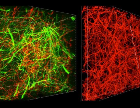
MAP is an advancement of a technique called CLARITY, both of which were developed by study author Kwanghun Chung. CLARITY allows researchers to see brain cells as transparent, so they can look inside them to examine the molecules that make them up.
MAP goes a step further than that, allowing for transparent viewing of the brain’s building blocks at multiple levels. The technique expands brain tissues up to four or five times their normal size without moving them around or compromising their structure.
To do that, researchers injected the brain with a gel made of acrylamide polymers, enlarging the cells to allow for clearer viewing, all while the structure of the cells, and the brain tissue itself, remains intact. Then, researchers introduce formaldehyde as a way to make brain proteins stick to the gel as the whole mixture expands. Next, the brain cells are injected with a fluorescent material to label and classify the molecules inside the cells.
With the molecules now lit up with fluorescent antibodies (different markers for different brain structures), researchers can use imaging technology to map it all out. They can see which brain molecules tend to group with others in what areas of the brain and as parts of which larger structures. This allows researchers to see which brain functions and brain materials are related to one another, which is important for understanding how the brain works as a whole.
The method isn’t constrained to the brain—other organs, such as the lungs, liver, kidneys and heart, could benefit from the intensely close-up imaging of MAP.
Read Full Article – Source: New technique allows for zooming in, out of brain imaging at different levels | Health Imaging
By: Caitlin Wilson